Immersion
Immersion Is… a unique opportunity for students to get hands-on research training and experiences related to neuroscience. Through this experiential learning, many students are exposed to new areas of science that they explore further, and even pursue as a career path.
Immersion Through Research
The neuroscience program is designed to provide students with immersive experiences in research. Students participate in research in the laboratories of neuroscience faculty under the auspices of the directed studies courses, NSC 3860, 3851, 3861/3862, and 3863/3864. Further research experience is available through the Honors Program in neuroscience.
Immersion Stories
Liyu “Leo” Huang, BA’24
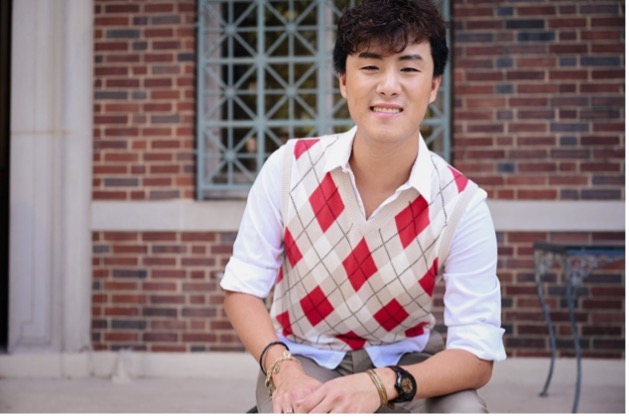 Leo, a triple major in neuroscience; architecture and the built environment; and medicine, health, and society, shaped an Immersion experience that used his artistic eye to help bridge communication between pathologists and surgeons in the operating room.
Alongside mentor Dr. Michael Topf, assistant professor of otolaryngology-head and neck surgery at Vanderbilt University Medical Center, Leo gained experience using 3D technology to scan tumor specimens being removed from patients.
Traditionally, in the operating room, a surgeon removes a tumor specimen from a patient and sends it off to the pathology lab. The pathologist then looks for “positive margins,” indicating that more of the tumor still needs to be removed and communicates that back to the surgeon. This process is normally done in 2D, with phone calls and anatomical references between the pathologist and the surgeon. Time is of the essence, as the patient is still under anesthesia in the operating room while this communication is happening.
In his research with Topf, Leo helped take 3D scans of tumor specimens that were removed from the patient. Consulting with the pathologist, Leo used a drawing tablet and CAD (computer-aided design) software to ensure that the colors and annotations provide an accurate visual representation of the specimens that can then be visually translated back to the operating room. This 3D technology improves communication between the pathologist and the surgeon about any positive margins and provides more accurate and timely results.
Read more and watch a video about Leo’s Immersion experience.